NBEO Part II (TMOD) · high-yield study reference

Corneal Dystrophies & Inherited Retinal Diseases

Your original chart, fact-checked against the IC3D 2024 classification, EyeWiki, EyeRounds and peer-reviewed reviews. Each condition now has a photo: slit lamp for the cornea, fundus for the retina. Notes in orange show what changed and link to the source. Click any photo to open its original page.

ADautosomal dominant ARautosomal recessive XLX-linked Mitomitochondrial (maternal) Sporadicusually not inherited
Shows only the photos. Tap a plate to reveal the answer.
32 changes from the original chart

Corneal dystrophies, anterior to posterior

Board rule of thumb: almost all corneal dystrophies are autosomal dominant. Memorize the exceptions: Macular (AR), Gelatinous drop-like (AR), CHED (AR, formerly CHED2) and Lisch (classically X-linked dominant; IC3D 2024 now lists it as AD).

Epithelial & subepithelial

Epithelial Basement Membrane (EBMD)

Map-dot-fingerprint / Cogan
Layer
Epithelium & basement membrane
Inheritance
Sporadicrare AD

Most common anterior corneal dystrophy; IC3D now considers it mostly degenerative. Geographic map lines, fingerprint lines, Cogan microcysts (dots). Causes recurrent corneal erosions (RCE). Negative staining with NaFl.

Changed from your original

  • Inheritance clarified: IC3D 2024 calls EBMD most likely degenerative; true AD families are rare. source
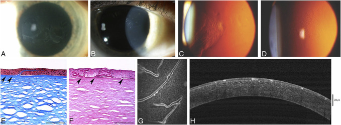
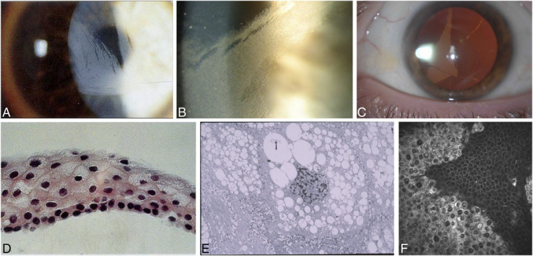
Slit lamp: IC3D Figure 1 (multi-panel). Use panels A-D: A, map-like changes (direct/diffuse illumination); B, intraepithelial dot opacities (Cogan microcysts) with maps; C, fingerprint lines on retroillumination; D, Bron bleb pattern on retroillumination. Panels E-H are histology, confocal and OCT.
Slit lamp IC3D Figure 1 (multi-panel). Use panels A-D: A, map-like changes (direct/diffuse illumination); B, intraepithelial dot opacities (Cogan microcysts) with maps; C, fingerprint lines on retroillumination; D, Bron bleb pattern on retroillumination. Panels E-H are histology, confocal and OCT.

Tap to reveal
Meesmann

Juvenile epithelial
Layer
Epithelium
Inheritance
AD

KRT3/KRT12. Myriad tiny intraepithelial microcysts, densest interpalpebrally and extending to the limbus; best seen on retroillumination. Usually asymptomatic or mild glare. Cysts contain PAS-positive debris; "peculiar substance" is the EM finding.

Changed from your original

  • "Peculiar substance" is an electron-microscopy finding; the PAS-positive material is the cyst debris on light microscopy. source
Slit lamp: Slit-lamp photo with broad-beam light: myriad tiny, uniform, round gray-white intraepithelial microcysts scattered across the cornea.
Slit lamp Slit-lamp photo with broad-beam light: myriad tiny, uniform, round gray-white intraepithelial microcysts scattered across the cornea.

Tap to reveal
Lisch Epithelial

Layer
Epithelium
Inheritance
XLDIC3D 2024: AD

Gray, feathery, whorled or band-shaped epithelial opacities; may or may not reach the limbus, can be asymmetric. Retroillumination shows densely crowded clear microcysts. Board answer is still usually X-linked dominant; IC3D 2024 reclassified it as AD (MCOLN1).

Changed from your original

  • Inheritance: IC3D 2024 reclassified Lisch as AD (MCOLN1) after the gene was found. Older references and EyeWiki still say XLD. source
  • Removed "interpalpebral zone" (that describes Meesmann). Reworded "clear in retroillumination" to "densely crowded clear microcysts on retroillumination". source
Slit lamp: IC3D Figure 5 (multi-panel). Use panels A-C: A and B, diffuse/direct illumination showing gray epithelial opacities in radial, feathery and club-shaped (whorled/band) patterns; C, retroillumination showing that the opacity consists of densely crowded transparent microcysts. Panels D-F are histology, EM and confocal.
Slit lamp IC3D Figure 5 (multi-panel). Use panels A-C: A and B, diffuse/direct illumination showing gray epithelial opacities in radial, feathery and club-shaped (whorled/band) patterns; C, retroillumination showing that the opacity consists of densely crowded transparent microcysts. Panels D-F are histology, EM and confocal.

Tap to reveal
Gelatinous Drop-Like

Layer
Subepithelial / anterior stroma
Inheritance
AR

Exception (AR), TACSTD2. Bilateral mulberry-like subepithelial amyloid nodules; severe photophobia and tearing early in life. Congo red positive.

Slit lamp: External/slit-lamp photo in diffuse white light: advanced GDLD with an almost entirely opaque cornea covered by multiple gelatinous, drop-like (mulberry) subepithelial amyloid nodules and some superficial vessels.
Slit lamp External/slit-lamp photo in diffuse white light: advanced GDLD with an almost entirely opaque cornea covered by multiple gelatinous, drop-like (mulberry) subepithelial amyloid nodules and some superficial vessels.

Tap to reveal
Bowman layer (TGFBI / 5q31)

Reis-Bücklers

CDB type I
Layer
Bowman layer & superficial stroma
Inheritance
AD

TGFBI R124L. Geographic or ring-like subepithelial opacities; early painful RCE and significant vision loss. Masson trichrome stains red. EM: rod-shaped bodies (same as granular type 1).

Changed from your original

  • Error fixed: the original said "curly fibers" on EM. Curly fibers belong to Thiel-Behnke; Reis-Bücklers shows rod-shaped bodies. source
Slit lamp: Slit-lamp photo in direct/diffuse white light: coarse reticular/geographic gray-white opacities at the level of Bowman layer and superficial stroma across the central cornea.
Slit lamp Slit-lamp photo in direct/diffuse white light: coarse reticular/geographic gray-white opacities at the level of Bowman layer and superficial stroma across the central cornea.

Tap to reveal
Thiel-Behnke

CDB type II
Layer
Bowman layer & superficial stroma
Inheritance
AD

TGFBI R555Q. Honeycomb-shaped subepithelial opacities; milder vision loss than Reis-Bücklers. Light microscopy (and AS-OCT): sawtooth pattern. EM: curly fibers (9–15 nm), the diagnostic finding.

Changed from your original

  • Error fixed: "sawtooth" is a light-microscopy pattern, not EM. The EM finding is curly fibers. source
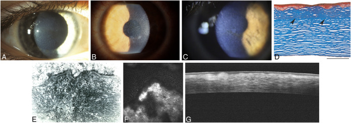
Slit lamp: IC3D Figure 8 (multi-panel). Use panels A-C: A, early mild honeycomb appearance (diffuse light); B, intense honeycomb subepithelial opacity in advanced disease (slit-lamp, broad diffuse light); C, honeycomb opacity in a 42-year-old with R555Q TGFBI. Panels D-G: light microscopy (sawtooth), EM (curly filaments), confocal and OCT.
Slit lamp IC3D Figure 8 (multi-panel). Use panels A-C: A, early mild honeycomb appearance (diffuse light); B, intense honeycomb subepithelial opacity in advanced disease (slit-lamp, broad diffuse light); C, honeycomb opacity in a 42-year-old with R555Q TGFBI. Panels D-G: light microscopy (sawtooth), EM (curly filaments), confocal and OCT.

Tap to reveal
Stroma · mnemonic: "Marilyn Monroe Always Gets Her Man In LA County"

Macular

Layer
Entire stroma, limbus to limbus, plus Descemet/endothelium
Inheritance
AR

Exception (AR), CHST6, and worst visual prognosis. Diffuse stromal haze with focal gray-white spots with indistinct borders; no clear spaces between opacities. Cornea is often thinner than normal. Mucopolysaccharides (GAGs) stain with Alcian blue (also colloidal iron).

Changed from your original

  • Added: corneal thinning (an omission, not an error). source
Slit lamp: Macular corneal dystrophy, slit lamp: discrete gray-white opacities within a diffusely hazy cornea with no clear intervening stroma.
Slit lamp Macular corneal dystrophy, slit lamp: discrete gray-white opacities within a diffusely hazy cornea with no clear intervening stroma.

Tap to reveal

Granular (Type 1)

Layer
Anterior / central stroma
Inheritance
AD

TGFBI R555W. Crumb-like, sharply demarcated white opacities with clear intervening stroma; spares the peripheral limbus. Best early VA. Hyaline stains red with Masson trichrome.

Slit lamp: Granular corneal dystrophy type 1: numerous discrete, irregular crumb-like white stromal opacities, central, sparing the limbus (advanced case, opacities partly confluent).
Slit lamp Granular corneal dystrophy type 1: numerous discrete, irregular crumb-like white stromal opacities, central, sparing the limbus (advanced case, opacities partly confluent).

Tap to reveal

Lattice (Type 1)

Layer
Anterior / central stroma
Inheritance
AD

TGFBI R124C. Refractile branching pipe-stem lines (best on retroillumination) plus recurrent corneal erosions. Amyloid stains orange-red with Congo red (apple-green birefringence). Old "type 2" (Meretoja) is now classed as systemic gelsolin amyloidosis, not a corneal dystrophy.

Changed from your original

  • Updated wording: Meretoja (old LCD type 2) is now familial systemic gelsolin amyloidosis (GSN), not a true corneal dystrophy. source
Slit lamp: Lattice corneal dystrophy: network of branching refractile linear stromal opacities. Note: this patient has a TGFBI L527R LCD variant (former 'LCD type III'), so the lines are thicker than in classic R124C LCD.
Slit lamp Lattice corneal dystrophy: network of branching refractile linear stromal opacities. Note: this patient has a TGFBI L527R LCD variant (former 'LCD type III'), so the lines are thicker than in classic R124C LCD.

Tap to reveal
Avellino

Granular type 2
Layer
Anterior / central stroma
Inheritance
AD

TGFBI R124H. Granular-type crumb, ring and stellate deposits plus short linear deposits (thicker and whiter than true lattice lines, rarely crisscross). Contains both hyaline and amyloid. LASIK/PRK contraindicated (worsens deposits).

Changed from your original

  • Reworded: IC3D discourages the old name "combined granular-lattice"; the linear deposits are not true lattice lines. source
Slit lamp: Granular corneal dystrophy type 2 (Avellino): variable-sized crumb-like and stellate stromal opacities, some fused into elongated linear shapes.
Slit lamp Granular corneal dystrophy type 2 (Avellino): variable-sized crumb-like and stellate stromal opacities, some fused into elongated linear shapes.

Tap to reveal

Schnyder

Crystalline
Layer
Central anterior stroma early, panstromal later
Inheritance
AD

UBIAD1. Central subepithelial needle-like cholesterol crystals (only ~50% of cases) plus prominent arcus lipoides plus panstromal haze. Associated with hypercholesterolemia.

Changed from your original

  • Layer corrected: starts centrally in the anterior stroma but becomes panstromal (the row already said "panstromal haze"). source
Slit lamp: Schnyder corneal dystrophy: central disc-shaped corneal opacity with fine subepithelial crystalline deposits.
Slit lamp Schnyder corneal dystrophy: central disc-shaped corneal opacity with fine subepithelial crystalline deposits.

Tap to reveal
Central Cloudy of François

Layer
Posterior stroma
Inheritance
SporadicAD?

Polygonal gray-white cloudy opacities separated by clear crack-like lines ("crocodile shagreen") in the deep central stroma. Asymptomatic; normal VA. IC3D: probably degenerative; indistinguishable from posterior crocodile shagreen.

Changed from your original

  • Inheritance nuance: IC3D says AD inheritance is reported but "difficult to substantiate"; most likely degenerative. source
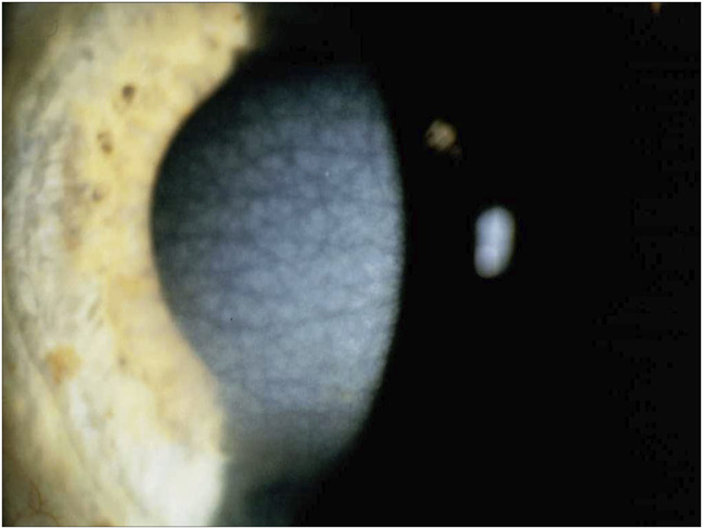
Slit lamp: IC3D Figure 19 (single panel): slit-lamp photo in diffuse/broad-beam light showing axially distributed, polygonal gray-white stromal opacities separated by linear areas of clear cornea (crocodile-shagreen pattern). IC3D notes no family history was available for this patient, so it may equally be posterior crocodile shagreen.
Slit lamp IC3D Figure 19 (single panel): slit-lamp photo in diffuse/broad-beam light showing axially distributed, polygonal gray-white stromal opacities separated by linear areas of clear cornea (crocodile-shagreen pattern). IC3D notes no family history was available for this patient, so it may equally be posterior crocodile shagreen.

Tap to reveal

Fleck

Layer
Stroma (all layers)
Inheritance
AD

PIKFYVE. Asymmetric (even unilateral), flat, gray-white dandruff-like specks throughout the stroma with clear intervening tissue. Usually asymptomatic with normal VA; occasionally mild photophobia or decreased corneal sensation.

Changed from your original

  • Corrected: the original said "normal corneal sensation"; IC3D and EyeWiki note sensation can be decreased. source
Slit lamp: Left (large) panel: slit-lamp optic section showing tiny discrete gray-white dandruff-like flecks scattered within the otherwise clear stroma. Right two panels: in vivo confocal microscopy (not slit lamp).
Slit lamp Left (large) panel: slit-lamp optic section showing tiny discrete gray-white dandruff-like flecks scattered within the otherwise clear stroma. Right two panels: in vivo confocal microscopy (not slit lamp).

Tap to reveal

Descemet membrane & endothelium

Fuchs Endothelial

Layer
Endothelium & Descemet membrane
Inheritance
ADSporadic

Most common endothelial dystrophy (F > M; TCF4 repeat). Central beaten-metal guttae, morning blurred vision, stromal edema, epithelial bullae. Tx: hypertonic NaCl, DSAEK/DMEK.

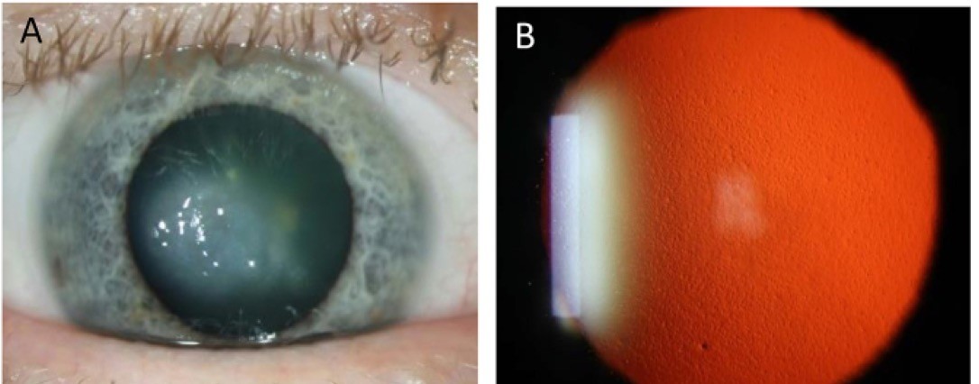
Slit lamp: Fuchs endothelial corneal dystrophy. Panel B: retroillumination showing numerous central guttae with a stippled 'beaten-metal' appearance. Panel A: central corneal edema. Cropped to panels A–B (histology and confocal panels removed).
Slit lamp Fuchs endothelial corneal dystrophy. Panel B: retroillumination showing numerous central guttae with a stippled 'beaten-metal' appearance. Panel A: central corneal edema. Cropped to panels A–B (histology and confocal panels removed).

Tap to reveal

Posterior Polymorphous (PPCD)

Layer
Endothelium & Descemet membrane
Inheritance
AD

ZEB1, OVOL2, GRHL2. Endothelial cells behave like epithelial cells (multilayered). Vesicles, railroad-track (snail-track) bands, geographic gray lesions; can cause iridocorneal adhesions and glaucoma.

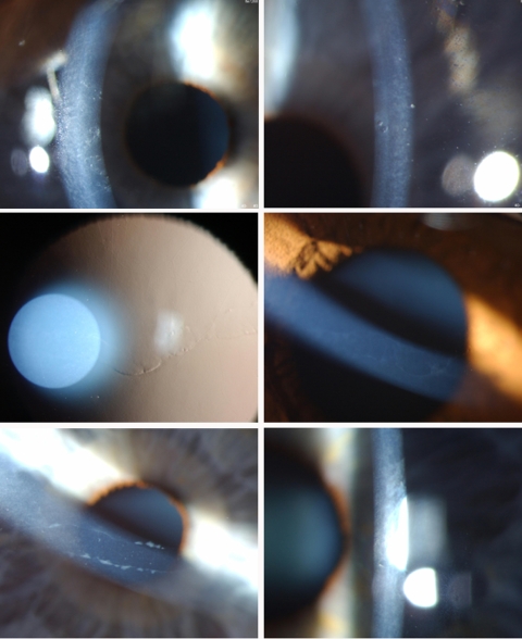
Slit lamp: PPCD slit-lamp phenotypes. Top row: scattered vesicular endothelial lesions. Middle row: retroillumination and direct view of a band lesion with a gray halo. Bottom row: band ('railroad-track') lesions with thickened Descemet edges. Low-resolution composite.
Slit lamp PPCD slit-lamp phenotypes. Top row: scattered vesicular endothelial lesions. Middle row: retroillumination and direct view of a band lesion with a gray halo. Bottom row: band ('railroad-track') lesions with thickened Descemet edges. Low-resolution composite.

Tap to reveal

Congenital Hereditary Endothelial (CHED)

Layer
Endothelium & Descemet membrane
Inheritance
AR

Exception (AR), SLC4A11. Bilateral (often asymmetric) diffuse ground-glass corneal clouding at birth or infancy with marked stromal thickening; thickened Descemet membrane; nystagmus common; IOP normal. CHED + deafness = Harboyan syndrome.

Changed from your original

  • Corrected: "bilateral symmetric" changed to "bilateral, often asymmetric" (IC3D). source
Slit lamp: CHED: diffuse, markedly opaque ground-glass cornea from stromal edema secondary to defective endothelium.
Slit lamp CHED: diffuse, markedly opaque ground-glass cornea from stromal edema secondary to defective endothelium.

Tap to reveal

Inherited retinal diseases, by primary layer

Board strategy: group retinal dystrophies by the layer where the primary defect sits, then lock in the inheritance, the hallmark fundus appearance and the ERG/EOG signature.

Photoreceptors (rods & cones)

Retinitis Pigmentosa (RP)

Layer
Photoreceptors (rods > cones)
Inheritance
ADARXLR

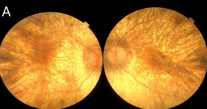
AD = mildest, latest onset; XLR = most severe, earliest onset. Classic triad: bone spicules, arteriolar attenuation, waxy disc pallor. Nyctalopia, ring scotoma. Scotopic ERG reduced and delayed (rod > cone) early, extinguished in advanced disease.

Changed from your original

  • Clarified: the scotopic ERG is reduced/delayed early and only becomes extinguished in advanced RP. source
Fundus: Mid-stage RP: bone-spicule pigment in the mid-periphery, attenuated retinal arterioles, and a relatively preserved macula with a ring of depigmentation around it.
Fundus Mid-stage RP: bone-spicule pigment in the mid-periphery, attenuated retinal arterioles, and a relatively preserved macula with a ring of depigmentation around it.

Tap to reveal

Cone-Rod Dystrophy

Layer
Photoreceptors (cones > rods)
Inheritance
ADARXL

Early loss of central VA, dyschromatopsia, hemeralopia (daylight glare). Bull's-eye maculopathy. Photopic/flicker ERG reduced first, followed by scotopic ERG.

Fundus: Cone-rod dystrophy in a 45-year-old with an ABCA4 loss-of-function variant. The posterior pole shows atrophy with pigment deposits of various shapes, while the periphery (top of the image) is less damaged. This is a later stage; early disease more often shows a bull's-eye maculopathy or subtle macular RPE change.
Fundus Cone-rod dystrophy in a 45-year-old with an ABCA4 loss-of-function variant. The posterior pole shows atrophy with pigment deposits of various shapes, while the periphery (top of the image) is less damaged. This is a later stage; early disease more often shows a bull's-eye maculopathy or subtle macular RPE change.

Tap to reveal

Achromatopsia

Rod monochromacy
Layer
Cone photoreceptors
Inheritance
AR

CNGA3, CNGB3. Absent/dysfunctional function of all 3 cone types. VA ~20/200, complete color blindness, severe photophobia, pendular nystagmus. Fundus normal or near-normal (OCT: ellipsoid-zone disruption). Absent photopic ERG, normal scotopic ERG.

Changed from your original

  • Nuance added: fundus is normal early but OCT usually shows ellipsoid-zone disruption; mild RPE changes can develop later. source
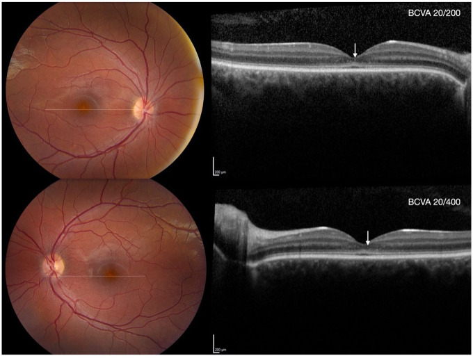
Fundus: CNGA3-related achromatopsia at baseline. Left: color fundus photos are near-normal apart from mild yellow foveal discoloration. Right: SD-OCT shows foveal outer-retinal change (flattened/disrupted ellipsoid zone, arrows). BCVA was 20/200 OD and 20/400 OS. The fundus is classically normal, so the OCT is the more useful image.
Fundus CNGA3-related achromatopsia at baseline. Left: color fundus photos are near-normal apart from mild yellow foveal discoloration. Right: SD-OCT shows foveal outer-retinal change (flattened/disrupted ellipsoid zone, arrows). BCVA was 20/200 OD and 20/400 OS. The fundus is classically normal, so the OCT is the more useful image.

Tap to reveal

Blue Cone Monochromacy

Layer
L & M cone photoreceptors
Inheritance
XLR

Only S-cones and rods work. VA 20/60–20/200, nystagmus, photophobia, often myopic; affected males. Only limited blue-yellow discrimination, mainly in dim light (rod + S-cone interaction); daylight color vision is essentially absent. S-cone ERG preserved.

Changed from your original

  • Corrected: the original said BCM "preserves blue-yellow discrimination". That overstates it; residual discrimination is limited and mostly mesopic. source
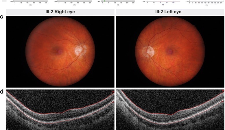
Fundus: Blue cone monochromacy in a 24-year-old man with a de novo deletion of the OPN1LW/OPN1MW gene cluster. (c) Fundus photos are essentially normal apart from tilted myopic discs. (d) OCT shows normal layering with thinned photoreceptor layers. Panels c-d are cropped from the full figure, which also shows ERG (a-b) and D-15 (e).
Fundus Blue cone monochromacy in a 24-year-old man with a de novo deletion of the OPN1LW/OPN1MW gene cluster. (c) Fundus photos are essentially normal apart from tilted myopic discs. (d) OCT shows normal layering with thinned photoreceptor layers. Panels c-d are cropped from the full figure, which also shows ERG (a-b) and D-15 (e).

Tap to reveal

Congenital Stationary Night Blindness (CSNB)

Layer
Photoreceptors / bipolar synapse
Inheritance
ADARXLR

Non-progressive nyctalopia from birth with a normal fundus. Schubert-Bornschein type (XL or AR; complete and incomplete) gives the electronegative ERG (normal a-wave, reduced b-wave). The AD Riggs type is not electronegative (reduced a- and b-waves).

Changed from your original

  • Corrected: only the Schubert-Bornschein forms (XL/AR) have an electronegative ERG; the AD Riggs type does not. source
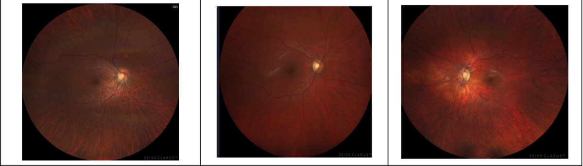
Fundus: CSNB fundi, which are essentially normal apart from myopic/tilted-disc changes. Left: Case 1, complete CSNB (autosomal recessive GPR179). Middle and right: Cases 2-3, brothers with X-linked incomplete CSNB (CACNA1F). Zeiss Clarus widefield. The fundus row is cropped from a figure that also shows OCT, mfERG and pVEP.
Fundus CSNB fundi, which are essentially normal apart from myopic/tilted-disc changes. Left: Case 1, complete CSNB (autosomal recessive GPR179). Middle and right: Cases 2-3, brothers with X-linked incomplete CSNB (CACNA1F). Zeiss Clarus widefield. The fundus row is cropped from a figure that also shows OCT, mfERG and pVEP.

Tap to reveal

Oguchi Disease

Layer
Rod photoreceptors
Inheritance
AR

Form of CSNB (SAG, GRK1). Golden-yellow metallic fundus sheen in light that disappears after prolonged (often 2–3 h) dark adaptation and returns within minutes of light (Mizuo-Nakamura phenomenon).

Changed from your original

  • Softened timing: reported dark-adaptation times range from ~45 min to 3+ h. source
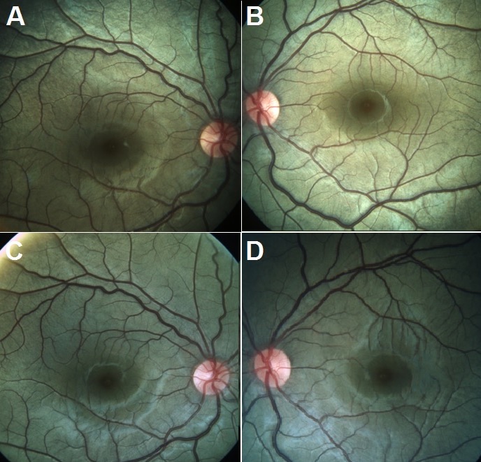
Fundus: Oguchi disease (Mizuo-Nakamura phenomenon) in a 7-year-old boy. (A, B) Light-adapted fundi show the golden-yellow (greenish-gold) metallic sheen. (C, D) After dark adaptation the sheen is gone and the fundi look normal. In this child it took about 45 min; it classically takes hours.
Fundus Oguchi disease (Mizuo-Nakamura phenomenon) in a 7-year-old boy. (A, B) Light-adapted fundi show the golden-yellow (greenish-gold) metallic sheen. (C, D) After dark adaptation the sheen is gone and the fundi look normal. In this child it took about 45 min; it classically takes hours.

Tap to reveal

Fundus Albipunctatus

Layer
Photoreceptors / RPE visual cycle
Inheritance
AR

Form of CSNB (RDH5). Numerous discrete yellow-white dots across the mid-periphery, sparing the macula; normal retinal vessels (unlike retinitis punctata albescens). Rod ERG and dark adaptation normalize after prolonged dark adaptation. Some older patients develop cone dystrophy.

Changed from your original

  • Added: rod ERG recovers after prolonged dark adaptation; not always stationary (some RDH5 patients develop cone dystrophy). source
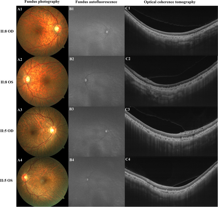
Fundus: Fundus albipunctatus in a Chinese family with RDH5 variants. The fundus photo column (A1-A4) shows numerous discrete yellow-white dots that spare the fovea, with normal-caliber vessels. The most striking panels are A3-A4 (patient II:5). The full figure also shows FAF (B) and OCT (C).
Fundus Fundus albipunctatus in a Chinese family with RDH5 variants. The fundus photo column (A1-A4) shows numerous discrete yellow-white dots that spare the fovea, with normal-caliber vessels. The most striking panels are A3-A4 (patient II:5). The full figure also shows FAF (B) and OCT (C).

Tap to reveal

Leber Congenital Amaurosis (LCA)

Layer
Photoreceptors & RPE
Inheritance
ARrare AD

Severe congenital blindness, roving nystagmus, sluggish pupils, Franceschetti oculodigital sign (eye poking), extinguished ERG. Voretigene neparvovec (Luxturna) gene therapy for biallelic RPE65 mutations.

Changed from your original

  • Clarified: mostly AR with rare AD forms (CRX, IMPDH1, OTX2); voretigene is only for biallelic RPE65. source
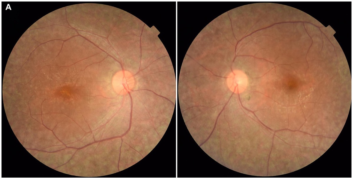
Fundus: RPE65-associated LCA in a 27-year-old Japanese man with infantile nystagmus and lifelong night blindness. Panel A (color fundus, OD left, OS right) shows diffuse RPE hypopigmentation/mottling and narrowed retinal vessels. The LCA fundus is very variable, from near-normal in infancy to RP-like.
Fundus RPE65-associated LCA in a 27-year-old Japanese man with infantile nystagmus and lifelong night blindness. Panel A (color fundus, OD left, OS right) shows diffuse RPE hypopigmentation/mottling and narrowed retinal vessels. The LCA fundus is very variable, from near-normal in infancy to RP-like.

Tap to reveal

Retinal pigment epithelium (RPE) & Bruch's membrane

Stargardt Disease

Fundus flavimaculatus
Layer
Photoreceptor outer segments (ABCA4) → lipofuscin in RPE
Inheritance
AR

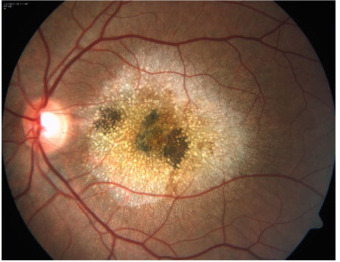
Most common juvenile macular dystrophy. ABCA4 defect in photoreceptor outer segments → lipofuscin (A2E) builds up in the RPE → secondary RPE and photoreceptor loss. Beaten-bronze macular atrophy plus yellow pisciform (fishtail) flecks. FA "dark/silent choroid" in up to ~60%.

Changed from your original

  • Layer clarified: ABCA4 is a photoreceptor protein; lipofuscin accumulates in the RPE. source
  • Dark choroid is not universal (up to ~62% of patients). source
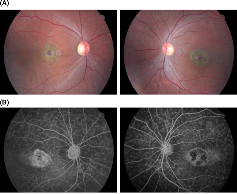
Fundus: Stargardt disease (compound heterozygous ABCA4), proband of a Han Chinese family. Top row: colour fundus photos (OD, OS) showing atrophic beaten-bronze macular lesion with surrounding yellowish flecks. Bottom row: fluorescein angiography showing hyperfluorescent flecks extending to the mid-periphery (Figure 2A-B of source; OCT panel C cropped out).
Fundus Stargardt disease (compound heterozygous ABCA4), proband of a Han Chinese family. Top row: colour fundus photos (OD, OS) showing atrophic beaten-bronze macular lesion with surrounding yellowish flecks. Bottom row: fluorescein angiography showing hyperfluorescent flecks extending to the mid-periphery (Figure 2A-B of source; OCT panel C cropped out).

Tap to reveal

Best Disease

Vitelliform macular dystrophy
Layer
RPE
Inheritance
AD

BEST1; incomplete penetrance and variable expressivity. Egg-yolk (vitelliform) yellow macular lesion in childhood, then pseudohypopyon, vitelliruptive and atrophic stages; VA often surprisingly good early. Diagnostic abnormal EOG (Arden ratio ≤ 1.5; normal ≥ 1.8) with normal full-field ERG.

Changed from your original

  • Arden cutoff written as ≤ 1.5 (normal ≥ 1.8); added incomplete penetrance. source
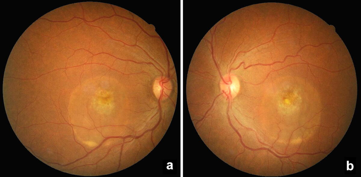
Fundus: Best vitelliform macular dystrophy (BEST1), proband of a Brazilian family. Colour fundus photographs of the right (a) and left (b) eye in the pseudohypopyon stage: yellow vitelliform material has settled inferiorly within the round macular lesion. Figure 2a-b of source; FAF, FA and OCT panels cropped out. This is not the classic intact 'egg-yolk' stage: no openly licensed, adequately sized egg-yolk-stage photo was found.
Fundus Best vitelliform macular dystrophy (BEST1), proband of a Brazilian family. Colour fundus photographs of the right (a) and left (b) eye in the pseudohypopyon stage: yellow vitelliform material has settled inferiorly within the round macular lesion. Figure 2a-b of source; FAF, FA and OCT panels cropped out. This is not the classic intact 'egg-yolk' stage: no openly licensed, adequately sized egg-yolk-stage photo was found.

Tap to reveal

Pattern Dystrophies

Butterfly, adult vitelliform
Layer
RPE
Inheritance
ADSporadic

PRPH2 most common (also BEST1, IMPG1/2, CTNNA1); many cases sporadic. Bilateral symmetric yellow/gray pigment figures (butterfly wings, reticular) at the macula; mild adult-onset vision loss. Normal ERG; EOG normal to mildly subnormal.

Changed from your original

  • PRPH2 is the most common gene, not the only one; EOG is normal to mildly subnormal depending on subtype. source
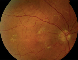
Fundus: Butterfly-shaped pattern dystrophy, right eye: colour fundus photo with yellowish butterfly-like flecks around the macula. Optic nerve head drusen, hard exudates and a secondary CNV are also present in this case. Figure 1A of source; the left-eye panel was cropped out. This is a modest, low-resolution example, not a textbook butterfly figure. EyeWiki's Pattern Dystrophies page has a clearer butterfly FAF image (copyrighted).
Fundus Butterfly-shaped pattern dystrophy, right eye: colour fundus photo with yellowish butterfly-like flecks around the macula. Optic nerve head drusen, hard exudates and a secondary CNV are also present in this case. Figure 1A of source; the left-eye panel was cropped out. This is a modest, low-resolution example, not a textbook butterfly figure. EyeWiki's Pattern Dystrophies page has a clearer butterfly FAF image (copyrighted).

Tap to reveal

Sorsby Fundus Dystrophy

Pseudoinflammatory
Layer
Bruch's membrane & RPE
Inheritance
AD

TIMP3. Thickened Bruch's membrane; early nyctalopia / delayed dark adaptation, then bilateral submacular CNV and disciform scarring in the 30s–40s.

Changed from your original

  • Added: night blindness and delayed dark adaptation are common first symptoms. source
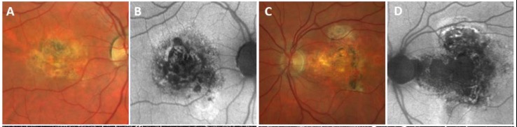
Fundus: Sorsby fundus dystrophy (TIMP3 p.Tyr152Cys), proband of Czech family 2. Panels A and C are colour fundus photos of the right and left eye: severe atrophy of the whole macular region with patchy pigmentation from long-standing CNV. Panels B and D are FAF showing central hypoautofluorescence. Top row of Figure 3; OCTA/OCT panels cropped out.
Fundus Sorsby fundus dystrophy (TIMP3 p.Tyr152Cys), proband of Czech family 2. Panels A and C are colour fundus photos of the right and left eye: severe atrophy of the whole macular region with patchy pigmentation from long-standing CNV. Panels B and D are FAF showing central hypoautofluorescence. Top row of Figure 3; OCTA/OCT panels cropped out.

Tap to reveal

Doyne Honeycomb

Malattia Leventinese
Layer
Bruch's membrane & RPE
Inheritance
AD

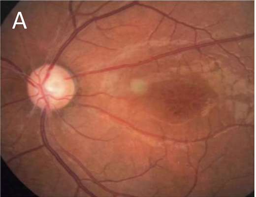
EFEMP1. Radial macular and peripapillary drusen arranged in a honeycomb pattern; drusen appear in the 20s–30s, symptoms usually in the 4th–5th decade; risk of secondary CNV.

Changed from your original

  • Clarified onset: drusen appear in the 20s–30s but symptoms usually start in the 4th–5th decade. source
Fundus: Malattia Leventinese / Doyne honeycomb retinal dystrophy (EFEMP1). Colour fundus photo of the left eye with confluent and radially arranged yellow-white drusen at the macula, extending to the peripapillary region, plus central pigmentary change and atrophy. Figure 1, panel B of source.
Fundus Malattia Leventinese / Doyne honeycomb retinal dystrophy (EFEMP1). Colour fundus photo of the left eye with confluent and radially arranged yellow-white drusen at the macula, extending to the peripapillary region, plus central pigmentary change and atrophy. Figure 1, panel B of source.

Tap to reveal

Inner retina (bipolar / Müller cells) & ganglion cell layer

X-Linked Juvenile Retinoschisis (XLRS)

Layer
Inner retina (Müller cells); foveal schisis mainly INL, peripheral schisis NFL
Inheritance
XLR

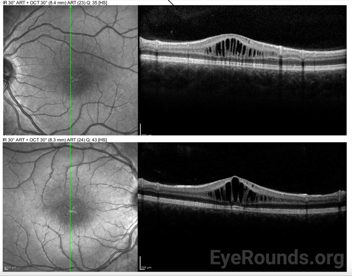
RS1 (retinoschisin). Spoke-wheel foveal schisis in nearly all patients (98–100%); peripheral schisis in ~50%, usually inferotemporal. Classic electronegative ERG (but a normal ERG doesn't rule it out). No leakage on FA.

Changed from your original

  • Corrected the layer: foveal schisis is mainly in the inner nuclear layer on OCT; splitting at the nerve fiber layer describes the peripheral schisis. source
  • "100%" changed to "nearly all"; "bullous" removed (bullous peripheral schisis is mainly seen in infants). source
Fundus: Color fundus photo, left eye, adult male with genetically confirmed XLRS (RS1): fine radial (spoke-wheel) striae around the fovea. Panel A of a figure, cropped. The disc cupping is from coexisting angle-closure glaucoma, not XLRS.
Fundus Color fundus photo, left eye, adult male with genetically confirmed XLRS (RS1): fine radial (spoke-wheel) striae around the fovea. Panel A of a figure, cropped. The disc cupping is from coexisting angle-closure glaucoma, not XLRS.
OCT: OCT through the fovea: cystoid schisis spaces mainly in the inner nuclear layer.
OCT OCT through the fovea: cystoid schisis spaces mainly in the inner nuclear layer.

Tap to reveal

Leber Hereditary Optic Neuropathy (LHON)

Layer
Retinal ganglion cells (RGCs)
Inheritance
Mito

Maternal inheritance; typically young males 15–35 (onset can be at any age). Subacute painless sequential bilateral central vision loss; circumpapillary telangiectatic microangiopathy and pseudoedema (no FA leak). mtDNA 11778 (ND4, most common, worst prognosis), 14484 (ND6, best recovery), 3460 (ND1). Tx: idebenone.

Changed from your original

  • Added the three primary mtDNA mutations and idebenone; noted that onset can occur outside 15–35. source
Fundus: Optic disc in acute LHON: a hyperemic, pseudo-edematous disc with circumpapillary telangiectatic microangiopathy (dilated, tortuous peripapillary vessels). Such a disc does not leak on FA.
Fundus Optic disc in acute LHON: a hyperemic, pseudo-edematous disc with circumpapillary telangiectatic microangiopathy (dilated, tortuous peripapillary vessels). Such a disc does not leak on FA.

Tap to reveal

Dominant Optic Atrophy (Kjer)

Layer
Retinal ganglion cells (RGCs)
Inheritance
AD

OPA1. Insidious bilateral symmetric central vision loss starting in the 1st–2nd decade; temporal (wedge-shaped) optic disc pallor, cecocentral scotoma, blue-yellow (tritan) color defect. Most patients stay better than 20/200.

Changed from your original

  • Onset widened from "first decade" to 1st–2nd decade (very insidious). source
Fundus: Fundus photos of the right (left image) and left (right image) eyes of a patient with an OPA1 mutation, showing optic disc pallor (arrows), most marked temporally. Caveat: this patient had an atypical acute, late-onset OPA1 presentation, so the pallor is more diffuse than the classic wedge-shaped temporal pallor of childhood-onset Kjer.
Fundus Fundus photos of the right (left image) and left (right image) eyes of a patient with an OPA1 mutation, showing optic disc pallor (arrows), most marked temporally. Caveat: this patient had an atypical acute, late-onset OPA1 presentation, so the pallor is more diffuse than the classic wedge-shaped temporal pallor of childhood-onset Kjer.

Tap to reveal

Choriocapillaris & choroid

Choroideremia

Layer
Choriocapillaris & RPE
Inheritance
XLR

CHM (REP-1). Progressive mid-peripheral atrophy of RPE and choriocapillaris exposing bare white sclera and large choroidal vessels; small central macular island preserved late. Female carriers show patchy peripheral pigment mottling.

Fundus: Color fundus photographs, right and left eyes, of a male with choroideremia: extensive atrophy of the RPE and choriocapillaris exposing large choroidal vessels and white sclera, with a relatively preserved central macular island and spared optic discs. Panel A of a multi-panel figure, cropped.
Fundus Color fundus photographs, right and left eyes, of a male with choroideremia: extensive atrophy of the RPE and choriocapillaris exposing large choroidal vessels and white sclera, with a relatively preserved central macular island and spared optic discs. Panel A of a multi-panel figure, cropped.

Tap to reveal

Gyrate Atrophy

Layer
Choriocapillaris & RPE
Inheritance
AR

Ornithine aminotransferase (OAT) deficiency → hyperornithinemia. Scalloped, sharply demarcated peripheral chorioretinal atrophy that coalesces toward the posterior pole; high myopia, early PSC cataract, CME. Tx: arginine-restricted diet; vitamin B6 for the B6-responsive minority.

Changed from your original

  • Clarified: vitamin B6 only helps the minority of patients who are B6-responsive. source
Fundus: Ultra-widefield color fundus images, right and left eyes: multiple sharply demarcated, scalloped, coalescing patches of chorioretinal atrophy in the mid to far periphery, with the posterior pole spared. Panel A of a multi-panel figure, cropped. In the source figure, panel C shows the associated CME.
Fundus Ultra-widefield color fundus images, right and left eyes: multiple sharply demarcated, scalloped, coalescing patches of chorioretinal atrophy in the mid to far periphery, with the posterior pole spared. Panel A of a multi-panel figure, cropped. In the source figure, panel C shows the associated CME.

Tap to reveal

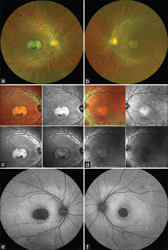
Central Areolar Choroidal Dystrophy

Layer
Photoreceptors & RPE, with secondary choriocapillaris atrophy
Inheritance
AD

PRPH2 (most often p.Arg142Trp). Well-circumscribed oval area of macular RPE and choriocapillaris atrophy exposing prominent choroidal vessels; no flecks or drusen. Changes start around age 20–40; serious vision loss usually in the 4th–7th decade.

Changed from your original

  • Added the gene (PRPH2). Layer corrected: histology shows loss of photoreceptors and RPE with secondary choriocapillaris atrophy. Onset range widened from 30–50. source
Fundus: Central areolar choroidal dystrophy. Panels a-b: widefield colour fundus photos. Panels c-d: multicolor images. Each shows a sharply demarcated round/oval atrophic area at the macula. Panels e-f: FAF showing hypoautofluorescence of the lesion, with no other abnormality. Full Figure 1 of source.
Fundus Central areolar choroidal dystrophy. Panels a-b: widefield colour fundus photos. Panels c-d: multicolor images. Each shows a sharply demarcated round/oval atrophic area at the macula. Panels e-f: FAF showing hypoautofluorescence of the lesion, with no other abnormality. Full Figure 1 of source.

Tap to reveal

Rapid-fire memory hooks

  • Electronegative ERG (normal a-wave, reduced b-wave): inner retina / bipolar dysfunction. Think X-linked retinoschisis and Schubert-Bornschein CSNB (complete and incomplete), plus CRAO, ischemic CRVO, melanoma-associated retinopathy and quinine toxicity. CRAO/CRVO are usually one eye; genetic causes are both.
  • Abnormal EOG with normal ERG: Best vitelliform macular dystrophy (Arden ratio ≤ 1.5; normal ≥ 1.8). Also abnormal in carriers with a normal-looking fundus.
  • X-linked recessive retinal trio: X-linked RP, X-linked retinoschisis and choroideremia (plus blue cone monochromacy and ocular albinism type 1, GPR143).
  • Stromal dystrophy stains ("Marilyn Monroe Always Gets Her Man In LA County"): Macular = Mucopolysaccharide / Alcian blue (AR); Granular = Hyaline / Masson trichrome (AD); Lattice = Amyloid / Congo red (AD).
Image credits